Posts

Showing posts from 2015
Image
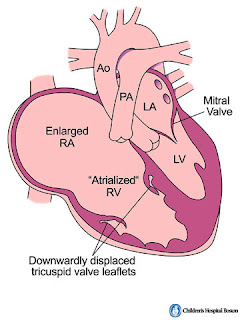
Ebstein's anomaly risk of anomaly 1 in 20,000 higher frequency in mothers who take lithium tricuspid valve leaflet anterior leaflet - largest leaflet and attached to valve annulus, large, redundant posterior and septal leaflet- vestigial/ absent - (failure of delamination of leaflets) when present the leaflets free edges are generally displaced posteriorly and downwards from AV junction and progressively towards the RVOT RV the RV i divided into two parts - proximal part - atrialized RV and distal part is RV proper and is small Different classifications to describe Ebstein's - type I--> IV (IV is the worst) Associated cardiac defects - ASD, PFO, VSD, PDA, RVOTO, Coarctation, WPW, LVNC Differential diagnosis of Ebstein's - tricuspid valve dysplasia - tricuspid valve prolapse - TV endocarditis - carcinoid heart disease - Uhl's anomaly - ARVC EKG findings RAE, RVH, RBBB, pre-excitation ECHO - TV displacement >0.8cm/m^2 of BSA com
Image
What is a Fontan? - palliative surgery which is performed in patients with a functional or an anatomic single ventricle - ventricle which is rudimentary or <30% of the expected volume. What are the typical anatomy for which a Fontan is performed? - HLHS - Tricusid atresia - Unbalanced AV canal - PA/IVS - DILV Rarely it may need to be performed in conditions such as - Ebstein's - DORV - ccTGA - any anatomy where there is severe hypoplasia of one of the ventricles Concept behind a fontan - there is a single ventricle and its job needs to be dedicated to pumping to the systemic circulation - therefore there is passive flow of blood through the Fontan - blood driven by central venous pressure - intra thoracic pressure changes - relaxation of the systemic ventricle 10 COMMANDMENTS FOR A SUCCESSFUL FONTAN PROCEDURE 1. Age - 2-7years of age 2. sinus rhythm 3. Normal systemic venous drainage 4. Normal right atrial volume 5. No/ minimal AVVR 6. Normal sing
Image
ECG interpretation in athletes ECG is very useful for early identification of causes of sudden cardiac death in athletes such as cardiomyopathy, primary electrical diseases. This interpretation becomes even more difficult in athletes where normal physiologic changes related to athletic training (Athlete's heart)  have to be distinguished from pathologic changes.  In 2012 a summit met at Seattle and put together a group of criteria to distinguish Athlete's heart form a pathological ECG findings (check out article at pubmed link http://www.ncbi.nlm.nih.gov/pubmed/23303758). The group discussed about 1. Normal ECG findings in athletes 2. Abnormal findings in cardiomyopathy 3. Abnormal findings in primary electrical  The above table is taken from the Seattle criteria guideline.  Check out the table below which gives you a great summary of abnormal ECG findings
Image
1 1/2 ventricular repair Biventricular repair ----> 1 1/2 ventricular repair ----> Single ventricle / Fontan pathway 1 1/2 repair - concept useful in case of borderline pulmonic ventricles. So the ventricle is not big enough to sustain a biventricular repair and is not small enough to commit the patient to a single ventricle route. Therefore this repair comprises of performing a Glenn procedure to offload the RV as well as intracardiac repair to keep open the future option of switching to a 2 ventricle pathway. http://www.ncbi.nlm.nih.gov/pubmed/9725452 Use the link above to access an excellent paper from Toronto by Dr. Freedom where the criteria for a 1 1/2 ventricle repair is discussed. This can be applied to 3 categories of patients 1. morphologic small pulmonary ventricle 2. functional deficits to the ventricle 3. special circumstances This table is taken directly from the article I was referring to from Toronto. The 1 1/2 ventricle repair has been fou
Image
Glenn with additional sources of pulmonary blood flow - what are the advantages? In some situations the Glenn anastomosis is performed along with an additional source of antegrade pulmonary blood flow, either with 1.  an additional systemic to PA shunt                          OR  2. Banding main pulmonary artery (Pulsatile Glenn). What are the advantages? 1. The pulmonary vascular bed is exposed to pulsatile pulmonary blood flow. According to certain studies exposing the pulmonary bed to this pulsatility improves growth of pulmonary vasculature pre-Fontan 2. Improved post operative SaO2. This effect may be negated in the long term where patients with only Glenn procedure may develop AP collaterals over time which causes their SaO2 to increase 3. The pulmonary vasculature is exposed to hepatic factor from the liver and therefore theoretically there is a lower likelihood of developing AP collaterals. What are the disadvantages? 1. The Glenn pressure is elevate